
File:Pulmonary embolism.jpg

本预览的尺寸:800 × 581像素。 其他分辨率:320 × 233像素 | 640 × 465像素 | 922 × 670像素。
原始文件 (922 × 670像素,文件大小:55 KB,MIME类型:image/jpeg)
| 描述 |
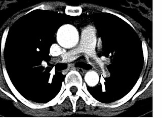
Čeština: CT - plicní embolie
English: Chest Spiral CT (with and without contrast agent) showing multiples filling defects of principal branches, due to acute and chronic pulmonary embolism. |
| 日期 | Published: 24 August 2007 |
| 来源 | Pulmonary embolism and patent foramen ovale thrombosis: the key role of TEE. Cardiovascular Ultrasound 2007, 5:26. doi:10.1186/1476-7120-5-26 |
| 作者 | Walter Serra, Giuseppe De Iaco, Claudio Reverberi and Tiziano Gherli |
| 授权 (二次使用本文件) |
文件历史
点击某个日期/时间查看对应时刻的文件。
| 日期/时间 | 缩略图 | 大小 | 用户 | 备注 | |
|---|---|---|---|---|---|
| 当前 | 2008年12月29日 (一) 16:09 | | 922 × 670(55 KB) | Stevenfruitsmaak | {{Information |Description={{en|1=Chest Spiral CT (with and without contrast agent) showing multiples filling defects of principal branches, due to acute and chronic pulmonary embolism.}} |Source=[http://www.cardiovascularultrasound.com/content/5/1/26 Pul |
文件用途
以下页面使用本文件:
全域文件用途
以下其他wiki使用此文件:
- ar.wikipedia.org上的用途
- az.wikipedia.org上的用途
- be.wikipedia.org上的用途
- es.wikipedia.org上的用途
- fr.wikipedia.org上的用途
- hi.wikipedia.org上的用途
- kk.wikipedia.org上的用途
- kn.wikipedia.org上的用途
- ru.wikipedia.org上的用途
- uk.wikipedia.org上的用途
- vi.wikipedia.org上的用途