
File:Chlamydomonas TEM 09.jpg

本预览的尺寸:751 × 600像素。 其他分辨率:301 × 240像素 | 601 × 480像素 | 961 × 768像素 | 1,280 × 1,023像素 | 1,800 × 1,438像素。
原始文件 (1,800 × 1,438像素,文件大小:784 KB,MIME类型:image/jpeg)
| 描述 |
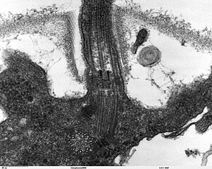
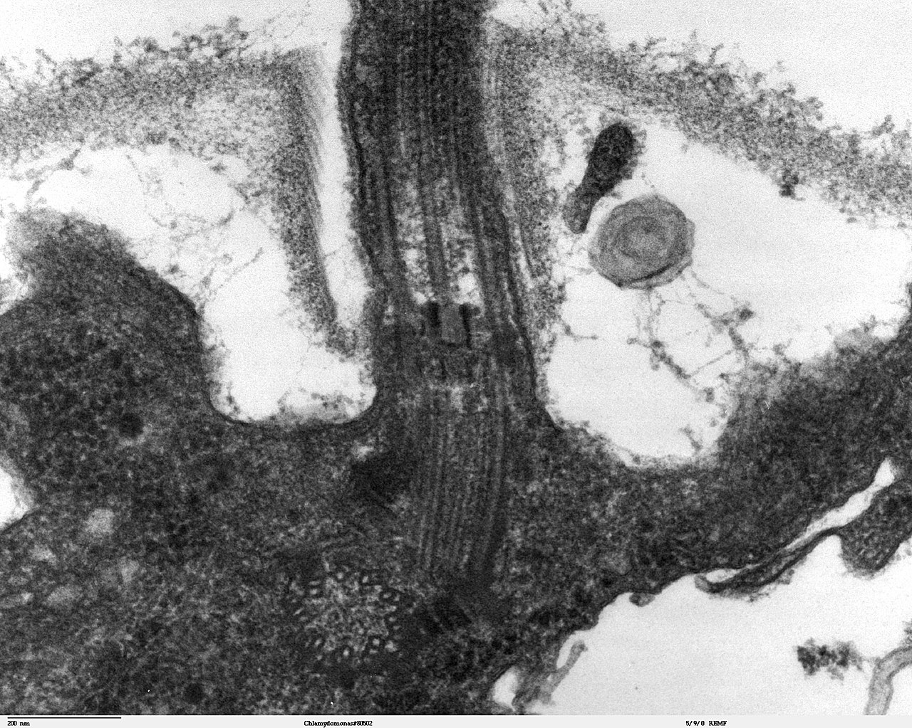
Transmission electron microscope image, showing an example of green algae (Chlorophyta). Chlamydomanas reinhardtii is a unicellular flagellate used as a model system in molecular genetics work and flagellar motility studies. This image is a longitudinal section through the flagella area. In the cell apex is the basal body that is the anchoring site for a flagella. Basal bodies originate from and have a substructure similar to that of centrioles, with nine peripheral microtubule triplets(see structure at bottom center of image). The two inner microtubules of each triplet in a basal body become the two outer doublets in the flagella. This image also shows the transition region, with its fibers of the stellate structure. The top of the image shows the flagella passing through the cell wall. |
| 日期 | |
| 来源 | Source and public domain notice at http://remf.dartmouth.edu/imagesindex.html |
| 作者 | Dartmouth Electron Microscope Facility, Dartmouth College |
| 授权 (二次使用本文件) |
Released into the public domain |
| 本作品已被作者Dartmouth Electron Microscope Facility, Dartmouth College释出到公有领域。这适用于全世界。 在一些国家这可能不合法;如果是这样的话,那么: Dartmouth Electron Microscope Facility, Dartmouth College无条件地授予任何人以任何目的使用本作品的权利,除非这些条件是法律规定所必需的。
|
文件历史
点击某个日期/时间查看对应时刻的文件。
| 日期/时间 | 缩略图 | 大小 | 用户 | 备注 | |
|---|---|---|---|---|---|
| 当前 | 2007年9月21日 (五) 06:47 | | 1,800 × 1,438(784 KB) | Neil916 | {{Information |Description= Transmission electron microscope image, showing an example of green algae (Chlorophyta). <br><br>''Chlamydomanas reinhardtii'' is a unicellular flagellate used as a model system in molecular genetics work and flagellar motilit |
文件用途
以下页面使用本文件:
全域文件用途
以下其他wiki使用此文件:
- ar.wikipedia.org上的用途
- bs.wikipedia.org上的用途
- ca.wikipedia.org上的用途
- cs.wikipedia.org上的用途
- de.wikipedia.org上的用途
- de.wikibooks.org上的用途
- en.wikipedia.org上的用途
- es.wikipedia.org上的用途
- gl.wikipedia.org上的用途
- id.wikipedia.org上的用途
- ja.wikipedia.org上的用途
- ko.wikipedia.org上的用途
- pl.wikipedia.org上的用途
- ru.wikipedia.org上的用途
- sv.wikipedia.org上的用途
- tr.wikipedia.org上的用途
- uk.wikipedia.org上的用途