
File:Birefringence microscopy of pseudogout, annotated.jpg
預覽大小:662 × 600 像素。 其他解析度:265 × 240 像素 | 530 × 480 像素 | 848 × 768 像素 | 1,130 × 1,024 像素 | 1,745 × 1,581 像素。
原始檔案 (1,745 × 1,581 像素,檔案大小:615 KB,MIME 類型:image/jpeg)
摘要
| 描述 |
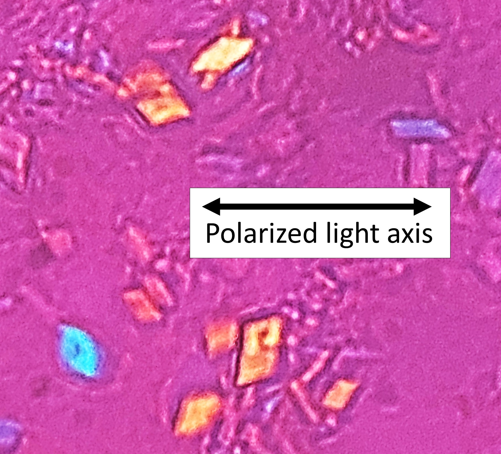
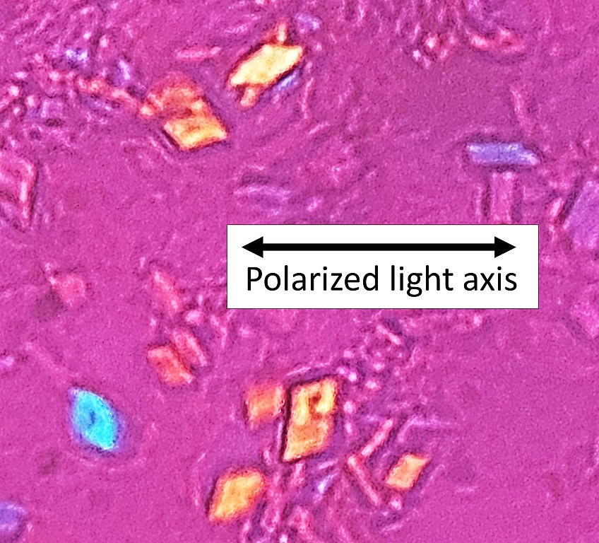
English: Microscopy with polarized light of tissue by a metatarsal joint, showing crystals whereof some (one annotated) have rhomboid shape and weak positive nirefringence, consistent with calcium pyrophosphate dihydrate crystal deposition disease (pseudogout). |
| 日期 | |
| 來源 | 自己的作品 |
| 作者 |
.jpg) - Reusing images - Conflicts of interest: None Consent note: Consent from the patient or patient's relatives is regarded as redundant, because of absence of identifiable features (List of HIPAA identifiers) in the media and case information (See also HIPAA case reports guidance). |
| 其他版本 |
 |
| 拍攝地點 | | 位於此地的本圖片與其他圖片: OpenStreetMap |
|---|
授權條款
| 此檔案在創用CC CC0 1.0 通用公有領域貢獻宣告之下分發。 | |
| 在此宣告之下分發本作品者,已依據各國著作權法,在全世界放棄其對本作品所擁有的著作權及所有相關相似的法律權利,從而將本作品貢獻至公有領域。您可以複製、修改、分發和演示該作品,用於任何商業用途,所有這些都不需要請求授權。
|
檔案歷史
點選日期/時間以檢視該時間的檔案版本。
| 日期/時間 | 縮圖 | 尺寸 | 使用者 | 備註 | |
|---|---|---|---|---|---|
| 目前 | 2022年4月4日 (一) 22:59 | | 1,745 × 1,581(615 KB) | Mikael Häggström | Sharper |
| 2020年11月12日 (四) 14:45 |  | 628 × 567(82 KB) | Mikael Häggström | +Axis | |
| 2020年11月12日 (四) 14:39 |  | 473 × 426(48 KB) | Mikael Häggström | Uploaded a work by {{Mikael Häggström|cat=Micrographs|consent=noid}} from {{Own}} with UploadWizard |
檔案用途
下列頁面有用到此檔案:
全域檔案使用狀況
以下其他 wiki 使用了這個檔案:
- ar.wikipedia.org 的使用狀況
- en.wikipedia.org 的使用狀況