
File:Leaf epidermis w scale.jpg

預覽大小:598 × 599 像素。 其他解析度:240 × 240 像素 | 479 × 480 像素 | 767 × 768 像素 | 1,022 × 1,024 像素 | 2,048 × 2,052 像素。
原始檔案 (2,048 × 2,052 像素,檔案大小:1.1 MB,MIME 類型:image/jpeg)
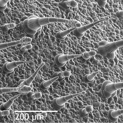
| 描述 | Scanning electron microscope image of Nicotiana alata upper leaf surface, showing tricomes and a few stomates. Instrument: ZEISS962 SEM. |
| 日期 | (UTC) |
| 來源 | |
| 作者 |
|
| 這是一張修飾過的圖片,即本圖片是用軟體修改過後的版本,修改的方式或內容有:Added scale, more contrast。原版圖片來源:Leaf epidermis.jpg。修改者:Laitr Keiows。
|
| 此作品已由其作者,Louisa Howard,釋出至公有領域。此授權條款在全世界均適用。 這可能在某些國家不合法,如果是的話: Louisa Howard授予任何人有權利使用此作品於任何用途,除受法律約束外,不受任何限制。
|
原始上傳日誌
This image is a derivative work of the following images:
- File:Leaf_epidermis.jpg licensed with PD-author
- 2008-06-21T18:26:19Z Mangostar 2048x2073 (3038992 Bytes) {{Information |Description=Scanning electron microscope image of Nicotiana alata upper leaf surface, showing tricomes and a few stomates. Instrument: ZEISS962 SEM. |Source=http://remf.dartmouth.edu/images/NicotianaLeafSEM/nic
Uploaded with derivativeFX
檔案歷史
點選日期/時間以檢視該時間的檔案版本。
| 日期/時間 | 縮圖 | 尺寸 | 使用者 | 備註 | |
|---|---|---|---|---|---|
| 目前 | 2010年3月11日 (四) 01:59 | | 2,048 × 2,052(1.1 MB) | Laitr Keiows | {{Information |Description=Scanning electron microscope image of Nicotiana alata upper leaf surface, showing tricomes and a few stomates. Instrument: ZEISS962 SEM. |Source=*File:Leaf_epidermis.jpg |Date=2010-03-11 01:58 (UTC) |Author=*[[:File:Leaf_e |
檔案用途
下列頁面有用到此檔案:
全域檔案使用狀況
以下其他 wiki 使用了這個檔案:
- ar.wikipedia.org 的使用狀況
- bn.wikipedia.org 的使用狀況
- bs.wikipedia.org 的使用狀況
- en.wikipedia.org 的使用狀況
- en.wikiversity.org 的使用狀況
- fr.wikipedia.org 的使用狀況
- gl.wikipedia.org 的使用狀況
- gv.wikipedia.org 的使用狀況
- ht.wikipedia.org 的使用狀況
- id.wikipedia.org 的使用狀況
- it.wikipedia.org 的使用狀況
- ja.wikipedia.org 的使用狀況
- kk.wikipedia.org 的使用狀況
- la.wikipedia.org 的使用狀況
- lv.wikipedia.org 的使用狀況
- ml.wikipedia.org 的使用狀況
- ms.wikipedia.org 的使用狀況
- ru.wikipedia.org 的使用狀況
- simple.wikipedia.org 的使用狀況
- sl.wikipedia.org 的使用狀況
- vi.wikipedia.org 的使用狀況