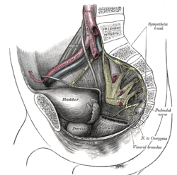
腰骶干
外观
| 腰骶干 | |
|---|---|
 腰骶神经丛解剖图 | |
 Dissection of side wall of pelvis showing sacral and pudendal plexuses. | |
| 基本信息 | |
| 来源 | L4-L5 |
| 标识字符 | |
| 拉丁文 | truncus lumbosacralis |
| TA98 | A14.2.07.026 |
| TA2 | 6504 |
| FMA | FMA:65535 |
| 格雷氏 | p.948 |
| 《神经解剖学术语》 [在维基数据上编辑] | |
腰骶干,一个连接腰神经丛与骶神经丛的神经组织,它是第四和第五腰椎神经的一部分共同形成的,并向下连接至骶神经丛。
解剖学
[编辑]腰骶干是从第五(L5)腰椎神经的整个前只和第四(L4)腰椎神经的一部分联合形成的。[1][2][3]L4 首先向腰神经丛发出分支,接下来从腰大肌[3]的内侧边缘出现,并与 L5 的前支在骨盆上部分略高于骨盆边缘处回合,形成粗后的的绳状干,[4]这穿越骨盆边缘(位于闭孔神经内侧)[3]后,下降到骶骨翼前表面,然后连接到骶神经丛[4]。
与骶神经一样,腰骶干分为前分支和后分支,然后重新合并形成下肢的屈肌和伸肌区域的神经。[3]
临床意义
[编辑]在分娩进入第二阶段的时候,胎儿的头部可能受到来自腰骶干的挤压,这可能导致一些腿部的肌肉无力[5],但是通常情况下是可以完全康复的。
更多照片
[编辑]-
腰骶干
-
骶交感神经与S1腰椎神经的连接
参考资料
[编辑]- ^ Mirjalili, S. Ali, Tubbs, R. Shane; Rizk, Elias; Shoja, Mohammadali M.; Loukas, Marios , 编, Chapter 46 - Anatomy of the Sacral Plexus L4-S4, Nerves and Nerve Injuries (San Diego: Academic Press), 2015-01-01: 619–626 [2021-01-13], ISBN 978-0-12-410390-0, (原始内容存档于2023-01-26) (英语)
- ^ Katirji, Bashar, Katirji, Bashar , 编, Case 5, Electromyography in Clinical Practice (Second Edition) (Philadelphia: Mosby), 2007-01-01: 117–124 [2021-01-13], ISBN 978-0-323-02899-8, (原始内容存档于2022-02-21) (英语)
- ^ 3.0 3.1 3.2 3.3 Sinnatamby, Chummy S. Last's Anatomy 12th. 2011: 310. ISBN 978-0-7295-3752-0.
- ^ 4.0 4.1 Moore, Keith L.; Dalley, Arthur F.; Agur, Anne M. R. Essential Clinical Anatomy. Lippincott Williams & Wilkins. 2017: 584. ISBN 978-1496347213.
- ^ Goyal, N.; Chad, D. A., Lumbar Plexopathy, Aminoff, Michael J.; Daroff, Robert B. (编), Encyclopedia of the Neurological Sciences (Second Edition), Oxford: Academic Press: 923–926, 2014-01-01 [2021-01-13], ISBN 978-0-12-385158-1, (原始内容存档于2022-02-21) (英语)
外部连结
[编辑]- 人体解剖学在线(Human Anatomy Online)网站上的相关图片:43:15-0103 - "The Female Pelvis: The Posterolateral Pelvic Wall"
- (英文)posteriorabdomen 在韦斯利诺曼的解剖课上(乔治城大学)
- figures/chapter_30/30-6.HTM — Basic Human Anatomy at Dartmouth Medical School


