股动脉
外观
| 股动脉 | |
|---|---|
 腹股沟韧带后方解剖 | |
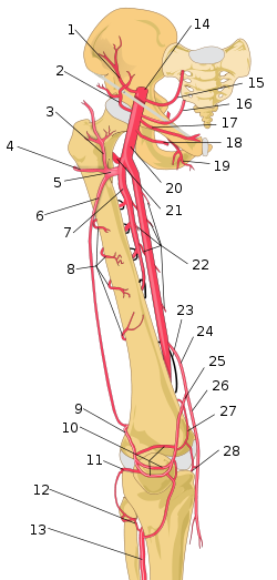
 Schema of femoral artery (labeled as #20) and its major branches - right thigh, anterior view. | |
| 基本信息 | |
| 来源 | 外髂动脉 |
| 分支 | 上腹壁动脉 浅旋髂动脉 浅外阴动脉 深外阴动脉 深股动脉 延续动脉为腘动脉 |
| 静脉 | 股静脉 |
| 供应范围 | 股前隔间 |
| 标识字符 | |
| 拉丁文 | Arteria femoralis |
| MeSH | D005263 |
| TA98 | A12.2.16.010 |
| TA2 | 4674 |
| FMA | FMA:70248 |
| 格雷氏 | p.623 |
| 《解剖学术语》 [在维基数据上编辑] | |
股动脉(拉丁语:arteria femoralis)是外髂动脉的直接延续,为供应下肢的主要动脉。 外髂动脉在经过腹股沟韧带深层后移行为总股动脉。之后总股动脉会分为股动脉及深股动脉,在股三角中沿前内侧下行。股动脉之后会穿过内收肌管,并进入内收大肌,进入内收大肌后股动脉移行为腘动脉[1]。
解剖构造
[编辑]
与股动脉相关的解剖构造如下:
- 前侧:股动脉前方被筋膜包围,位于缝匠肌后方。
- 后侧:股动脉由起源处往肢端分别枕在髂腰肌、耻骨肌,和内收长肌上方,股静脉则在股动脉和内收长肌间伴行。
- 内侧:股静脉仔股动脉上端延内侧下行。
- 外侧:股神经及其分支。
下列是股动脉的分支:
- 浅旋髂动脉:为股动脉的一条小分支,会上行至前上髂嵴。
- 浅腹壁动脉:为股动脉的一条小分支,会跨越腹股沟韧带上行至肚脐。
- 浅外阴动脉:为股动脉的一条小分支,会向内行走至阴囊(或大阴唇),供应该处表皮。
- 深外阴动脉:为股动脉的一条小分支,会向内行走至阴囊(或大阴唇),供应该处表皮。
- 股深动脉为股动脉相当重要的一条大分支,股深动脉会由腹股沟韧带下方4公分处,自股动脉外侧发出,会再其他大腿血管后方向内行走,并进入大腿内隔间。股深动脉会分出四条穿通支,最后一条穿通支唯股深动脉的终末分支。股深动脉在起点处有有两条分支,分别为内旋股动脉和外旋股动脉。
- 膝降动脉位于股动脉末端,接近股动脉将穿入内收大孔处,该动脉供应膝关节。
在临床上,股动脉未分支出股深动脉以前又常被称为“股总动脉”(Common femoral artery)而股动脉的延续分支则又被称做股浅动脉(superficial femoral artery)[2] 或缝下动脉 (subsartorial artery)[3]。
参考文献
[编辑]- ^ Schulte, Erik; Schumacher, Udo. Arterial Supply to the Thigh. Ross, Lawrence M.; Lamperti, Edward D. (编). Thieme Atlas of Anatomy: General Anatomy and Musculoskeletal System. Thieme. 2006: 490 [2015-11-26]. ISBN 978-3-13-142081-7. (原始内容存档于2021-03-08).
- ^ Richard .S. Snell (2008), Clinical Anatomy By Regions, 8th edition, Lippincott Williams & Wilkins, Baltimore, pages 581-582. http://books.google.co.zw/books?id=7SZWRe2OBlgC&pg=PA659&dq=clinical+anatomy+by+regions+lower+limb+femoral+artery&hl=en&sa=X&ei=3jQJVJT3Fs_daI-rgLgO&ved=0CBsQ6AEwAA (页面存档备份,存于互联网档案馆)
- ^ Amarnath C and Hemant Patel. Comprehensive Textbook of Clinical Radiology - Volume III: Chest and Cardiovascular system. [Elsevier Health Sciences. 2023 [2023-08-20]. ISBN 9788131263617. (原始内容存档于2023-08-16). Page 1072 (页面存档备份,存于互联网档案馆)
参见
[编辑]上臂动脉与股动脉作用类似。
本条目使用了部分解剖术语。
其他图像
[编辑]-
Structures surrounding right hip-joint.
-
Femoral sheath laid open to show its three compartments.
-
The femoral artery.
-
Segments of the femoral artery.[1]
-
The spermatic cord in the inguinal canal.
-
Front of right thigh, showing surface markings for bones, femoral artery and femoral nerve.
-
Schema of arteries of the thigh.
-
Femoral artery and its major branches - right thigh, anterior view.
-
Illustration depicting main leg arteries (anterior view).
-
Femoral artery.Deep dissection.
-
Femoral artery.Deep dissection.
外部链接
[编辑]- 人体解剖学在线(Human Anatomy Online)网站上的相关图片:12:05-0101
- 横截面图像:pelvis/pelvis-e12-15 - 维也纳医科大学生物塑化实验室提供
- Image at umich.edu - pulse
- Diagram at MSU
- ^ 引用错误:没有为名为
Amarnath2023的参考文献提供内容


![Segments of the femoral artery.[1]](http://upload.wikimedia.org/wikipedia/commons/thumb/4/46/Common_femoral_and_subsartorial_artery_and_vein.jpg/120px-Common_femoral_and_subsartorial_artery_and_vein.jpg)


