腹部超聲波
| 腹部超聲波檢查 | |
|---|---|
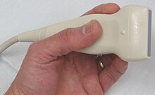
 腹部超聲波檢查所用的超聲波掃描儀 | |
| ICD-9-CM | 88.76 |
| OPS-301 | 3-059 |
| MedlinePlus | 003777 |
腹部超聲波是一種醫學超聲波檢查,用於檢查人體腹部。在病人腹部塗上超聲波導電凝膠後,超聲波探頭發出的聲波就能穿透腹部表面的腹壁,到達各器官。超聲波探頭將從器官反射的超聲波收集分析後,便能產出腹部影像。因此,這項檢查也被稱為跨腹超聲波,與直接將探頭以內窺鏡方式放入人體中空器官的內鏡超聲波有所不同。
腹部超聲波檢查通常由腸胃科、內科或放射科醫生進行,亦可由超聲波技師進行。
用途
[編輯]
腹部超聲波可用於判斷不同體內器官的異常,例如腎臟[1]、肝臟、膽囊、胰臟、脾臟、腹主動脈等。如果超聲波儀器有多普勒超聲波功能,還可以檢查血管中的血液流動狀況,協助診斷腎動脈狹窄等疾病。此外,腹部超聲波亦常用於檢查懷孕婦女的子宮與胎兒;這類檢查稱為產科超音波[2][3]。
當病患出現腹痛或急性腹痛,腹部超聲波可用於診斷病者是否患上闌尾炎及膽囊炎,以便安排緊急手術[4][5]。
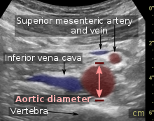
當醫生懷疑腹部器官可能異常脹大時,也會進行腹部超聲波檢查。能發現的病症包括腹主動脈瘤、脾臟腫大、尿瀦留等。診斷為腹主動脈瘤的標準是:腹主動脈(以最外層計算)的直徑超過3厘米,即為腹主動脈瘤[6]。
脾臟腫大是傳染性單核白血球增多症的常見症狀。腹部超聲波可以協助檢查患上此症者的狀況[7],但由於正常人體脾臟的大小差異很大,超聲波只應用於協助診斷脾臟腫大,而不應作為診斷的唯一依據,也不應僅依據超聲波結果來決定病者是否適合恢復運動[7]。
腹部超聲波也用於檢查腎臟功能異常、胰臟消化酶(如澱粉酶、胰臟脂酶)功能異常的病人。

結石檢查
[編輯]超聲波能發現體內的結石,包括腎石、膽結石等。由於結石會吸收超聲波,影像上將看到結石的後方出現黑色陰影。[8]
超聲波亦可用於導引不同治療程序,如導引體外震波、針刺活檢及腹部穿刺引流(通過針刺從腹腔中抽走積水的治療)等等[9]。
肝臟
[編輯]
腹部超聲波有助診斷肝功能指數異常的原因。超聲波圖像中可以看到的異常包括肝腫大[11]、反射增強(可能由膽汁鬱積所致)[12]、膽囊或膽管病症、肝臟腫瘤等[13]。
腎臟超聲波
[編輯]
腎臟超聲波是診斷與跟進腎臟疾病的重要常用工具。腎臟超聲波的圖像清晰,而且大部分腎臟病變都能在超聲波圖像中識別出來。[14]
技術特點
[編輯]腹部超聲波的優點包括:方便快捷、可直接在病床邊進行檢查、不需使用對人體(特別是孕婦)有危害的X光、相比其他腹部造影檢查(如電腦掃描)便宜等等[15]。但一個主要缺點則是,如果病人腸道內有大量氣體,或腹部脂肪較多,將難以進行檢查,影像的品質也不好[16]。此外,檢查中能否獲取滿意的超聲波影像,相當依賴進行檢查的醫護人員的經驗與技術水平[17]。
腹部超聲波的影像在檢查時就可以即時看到[18],進行檢查時也不須麻醉,所以可以通過移動探頭來檢查病人的反應[19]。例如,將探頭按在病人的膽囊上,如果病人感到痛楚,即可能是患上急性膽囊炎[20]。
超聲波能夠穿透腹壁,檢查骨盆內的器官與組織,例如膀胱、卵巢、子宮等。水是超聲波極佳的傳導媒介,所以檢查這些器官前,會請病人大量喝水,讓膀胱盡量脹大,以便超聲波訊號穿透[21][22]。
參考資料
[編輯]- ^ Bisset. Differential Diagnosis in Abdominal Ultrasound, 3/e. Elsevier India. 2008-01-01: 257 [2011-04-10]. ISBN 978-81-312-1574-6.
- ^ Whitworth, M; Bricker, L; Mullan, C. Ultrasound for fetal assessment in early pregnancy. Cochrane Database of Systematic Reviews. 2015, (7): CD007058. PMC 4084925
 . PMID 26171896. doi:10.1002/14651858.CD007058.pub3.
- ^ Salomon, LJ; Alfirevic, Z; Bilardo, CM; Chalouhi, GE; Ghi, T; Kagan, KO; Lau, TK; Papageorghiou, AT; Raine-Fenning, NJ; Stirnemann, J; Suresh, S; Tabor, A; Timor-Tritsch, IE; Toi, A; Yeo, G. ISUOG Practice Guidelines: performance of first-trimester fetal ultrasound scan (PDF). Ultrasound Obstet Gynecol. 2013, 41: 102–113 [2015-05-12]. PMID 23280739. doi:10.1002/uog.12342
 . (原始內容 (PDF)存檔於2015-09-06).
- ^ Puylaert, Julien B.C.M.; Rutgers, Peter H.; Lalisang, Roy I.; de Vries, Bas C.; van der Werf, Sjoerd D.J.; Dörr, Joep P.J.; Blok, Roeland A.P.R. A Prospective Study of Ultrasonography in the Diagnosis of Appendicitis. New England Journal of Medicine. 1987-09-10, 317 (11): 666–669. ISSN 0028-4793. PMID 3306375. doi:10.1056/NEJM198709103171103.
- ^ Ultrasonography by emergency physicians in patients with suspected cholecystitis. The American Journal of Emergency Medicine. 2001-01-01, 19 (1): 32–36 [2021-09-13]. ISSN 0735-6757. doi:10.1053/ajem.2001.20028. (原始內容存檔於2021-09-13) (英語).
- ^ 6.0 6.1 Timothy Jang. Bedside Ultrasonography Evaluation of Abdominal Aortic Aneurysm - Technique. Medscape. 2017-08-28.
- ^ 7.0 7.1 American Medical Society for Sports Medicine, Five Things Physicians and Patients Should Question, Choosing Wisely: an initiative of the ABIM Foundation (American Medical Society for Sports Medicine), 24 April 2014 [29 July 2014], (原始內容存檔於2014-07-29), which cites
- Putukian, M; O'Connor, FG; Stricker, P; McGrew, C; Hosey, RG; Gordon, SM; Kinderknecht, J; Kriss, V; Landry, G. Mononucleosis and athletic participation: an evidence-based subject review. Clinical Journal of Sport Medicine. 2008-07, 18 (4): 309–15. PMID 18614881. doi:10.1097/JSM.0b013e31817e34f8.
- Spielmann, AL; DeLong, DM; Kliewer, MA. Sonographic evaluation of spleen size in tall healthy athletes.. AJR. American Journal of Roentgenology. 2005-01, 184 (1): 45–9. PMID 15615949. doi:10.2214/ajr.184.1.01840045.
- ^ Dunmire, Barbrina; Harper, Jonathan D.; Cunitz, Bryan W.; Lee, Franklin C.; Hsi, Ryan; Liu, Ziyue; Bailey, Michael R.; Sorensen, Mathew D. Use of the Acoustic Shadow Width to Determine Kidney Stone Size with Ultrasound. Journal of Urology. 2016-01, 195 (1): 171–177 [2021-09-12]. doi:10.1016/j.juro.2015.05.111.
- ^ Dogra, Vikram.; Saad, Wael E. A. Ultrasound-guided procedures. New York, NY: Thieme. 2010. ISBN 9781604061703.
- ^ Christoph F. Dietrich; Carla Serra; Maciej Jedrzejczyk. Ultrasound of the liver - EFSUMB – European Course Book (PDF). European federation of societies for ultrasound in medicine and biology (EFSUMB). 2010-07-28 [2017-12-22]. (原始內容 (PDF)存檔於2017-08-12).
- ^ Childs, Jessie T; Esterman, Adrian J; Thoirs, Kerry A; Turner, Richard C. Ultrasound in the assessment of hepatomegaly: A simple technique to determine an enlarged liver using reliable and valid measurements: Ultrasound in the assessment of hepatomegaly. Sonography. 2016-06, 3 (2): 47–52 [2021-09-13]. doi:10.1002/sono.12051. (原始內容存檔於2021-09-13).
- ^ Di Serafino, Marco; Gioioso, Matilde; Severino, Rosa; Esposito, Francesco; Vezzali, Norberto; Ferro, Federica; Pelliccia, Piernicola; Caprio, Maria Grazia; Iorio, Raffaele; Vallone, Gianfranco. Ultrasound findings in paediatric cholestasis: how to image the patient and what to look for. Journal of Ultrasound. 2020-03, 23 (1): 1–12 [2021-09-13]. doi:10.1007/s40477-019-00362-9. (原始內容存檔於2022-03-14).
- ^ Janice Hickey, Franklin Goldberg. Ultrasound review of the abdomen, male pelvis & small parts. Philadelphia : Lippincott. 1999. ISBN 0397516916.
- ^ 內容來自: Hansen, Kristoffer; Nielsen, Michael; Ewertsen, Caroline. Ultrasonography of the Kidney: A Pictorial Review. Diagnostics. 2015, 6 (1): 2. ISSN 2075-4418. PMC 4808817
 . PMID 26838799. doi:10.3390/diagnostics6010002. (CC-BY 4.0) (頁面存檔備份,存於網際網路檔案館)
- ^ Noone, Tara C.; Semelka, Richard C.; Chaney, Deneise M.; Reinhold, Caroline. Abdominal imaging studies: comparison of diagnostic accuracies resulting from ultrasound, computed tomography, and magnetic resonance imaging in the same individual. Magnetic Resonance Imaging. 2004-01, 22 (1): 19–24 [2021-09-12]. doi:10.1016/j.mri.2003.01.001. (原始內容存檔於2021-09-12).
- ^ Berthold Block. Abdominal Ultrasound: Step by Step 2nd edition. Georg Thieme Verlag, Stuttgart, Germany: Thieme. 2012 [2021-09-12]. ISBN 9783131383631. (原始內容存檔於2022-08-07).
- ^ Miele, Vittorio; Piccolo, Claudia Lucia; Galluzzo, Michele; Ianniello, Stefania; Sessa, Barbara; Trinci, Margherita. Contrast-enhanced ultrasound (CEUS) in blunt abdominal trauma. The British Journal of Radiology. 2016-05, 89 (1061): 20150823 [2021-09-12]. doi:10.1259/bjr.20150823. (原始內容存檔於2021-09-12).
- ^ García de Casasola Sánchez, G.; Torres Macho, J.; Casas Rojo, J.M.; Cubo Romano, P.; Antón Santos, J.M.; Villena Garrido, V.; Diez Lobato, R. Abdominal Ultrasound and Medical Education. Revista Clínica Española (English Edition). 2014-04, 214 (3): 131–136 [2021-09-12]. doi:10.1016/j.rceng.2013.11.001. (原始內容存檔於2021-09-12).
- ^ Access to Assistive Technology and Medical Devices, Access to Medicines and Health Products, Health Product Policy and Standards, Medical Devices and Diagnostics. Elisabetta Buscarini, Harald Lutz and Paoletta Mirk , 編. Manual of diagnostic ultrasound 2nd ed. World Health Organization. 2013 [2021-09-12]. ISBN 9789241548540. (原始內容存檔於2022-03-20).
- ^ Simeone, Jf; Brink, Ja; Mueller, Pr; Compton, C; Hahn, Pf; Saini, S; Silverman, Sg; Tung, G; Ferrucci, Jt. The sonographic diagnosis of acute gangrenous cholecystitis: importance of the Murphy sign. American Journal of Roentgenology. 1989-02, 152 (2): 289–290 [2021-09-12]. PMID 2643262. doi:10.2214/ajr.152.2.289. (原始內容存檔於2022-08-10).
- ^ Benacerraf, B R; Shipp, T D; Bromley, B. Is a full bladder still necessary for pelvic sonography?. Journal of Ultrasound in Medicine. 2000-04, 19 (4): 237–241 [2021-09-12]. doi:10.7863/jum.2000.19.4.237. (原始內容存檔於2021-09-12).
- ^ NHS. Ultrasound scan. nhs.uk. 2017-10-18 [2021-09-12]. (原始內容存檔於2022-10-19) (英語).
