Lateral surface of left cerebral hemisphere, viewed from above. Angular gyrus is shown in orange.
Lateral surface of left cerebral hemisphere, viewed from the side. Angular gyrus is shown in orange.
Lateral view of a human brain, main gyri labeled.
Cerebrum. Lateral view.Deep dissection.
Cerebrum. Lateral view.Deep dissection.
Cerebrum. Lateral view.Deep dissection.
參考文獻
^Park, HJ; Kim, JJ; Lee, SK; Seok, JH; Chun, J; Kim, DI; et al. Corpus callosal connection mapping using cortical gray matter parcellation and DT-MRI. Human Brain Mapp. 2008, 29 (5): 503–16. PMID 17133394. doi:10.1002/hbm.20314.
^Makris, Nikos; Kennedy, David N.; McInerney, Sean; Sorensen, A. Gregory; Wang, Ruopeng; Verne, S. Caviness Jr; Pandya, Deepak N. Segmentation of Subcomponents within the Superior Longitudinal Fascicle in Humans: A Quantitative, In Vivo, DT-MRI Study. Cereb. Cortex. 2005, 15 (6): 854–869. PMID 15590909. doi:10.1093/cercor/bhh186.
^ 3.03.1Uddin, Lucina Q.; Supekar, Kaustubh; Amin, Hitha; Rykhlevskaia, Elena; Nguyen, Daniel A.; Greicius, Michael D.; Menon, Vinod. Dissociable Connectivity within Human Angular Gyrus and Intraparietal Sulcus: Evidence from Functional and Structural Connectivity. Cereb. Cortex. 2010, 20 (11): 2636–2646. doi:10.1093/cercor/bhq011.
^Rushworth, MF; Behrens, TE; Johansen-Berg, H. Connection patterns distinguish 3 regions of human parietal cortex. Cereb Cortex. 2006, 16: 1418–1430. doi:10.1093/cercor/bhj079.
^Makris, Nikos; Papadimitriou, George M.; Sorg, Scott; Kennedy, David N.; Caviness, Verne S.; Pandya, Deepak N. The occipitofrontal fascicle in humans: A quantitative, in vivo, DT-MRI study. NeuroImage. 2007, 37 (4): 1100–1111. doi:10.1016/j.neuroimage.2007.05.042.


 Position of angular gyrus (shown in red).
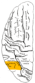 Lateral surface of left cerebral hemisphere, viewed from above. Angular gyrus is shown in orange.
 Lateral surface of left cerebral hemisphere, viewed from the side. Angular gyrus is shown in orange.
 Lateral view of a human brain, main gyri labeled.
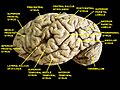 Cerebrum. Lateral view.Deep dissection.
 Cerebrum. Lateral view.Deep dissection.
 Cerebrum. Lateral view.Deep dissection.


