
滤泡旁细胞
| 滤泡旁细胞 | |
|---|---|
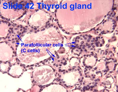
 Microscopic section of the thyroid showing follicles, where parafollicular cells reside | |
| 基本信息 | |
| 位置 | 甲状腺 |
| 功能 | 分泌降钙素 |
| 标识字符 | |
| TH | H3.08.02.4.00009 |
| FMA | FMA:68653 |
| 《显微解剖学术语》 [在维基数据上编辑] | |
滤泡旁细胞(parafollicular cell)又称C细胞,为人类及大部分脊椎动物甲状腺的一种细胞,胚胎学上源自于外胚层衍生之神经脊,为分泌降钙素的神经内分泌细胞[1]。滤泡旁细胞通常驻留于滤泡和滤泡上皮细胞的结缔组织间,因而得名。正常甲状腺切片中滤泡旁细胞通常不明显,但在某些发炎状况下会因增生而在苏伊染色(HE染色)下可见[2]。滤泡旁细胞在切片下可见时,通常比滤泡细胞大,细胞质较多,看起来有苍白的色斑,周围有胶状物围绕。
滤泡旁细胞在苏伊染色切片中,细胞质(胞质)稍淡;而用镀银法(银染法)可见基底部胞质内有嗜银颗粒,颗粒内有降钙素;电子显微镜下,位于滤泡上皮细胞之间的滤泡旁细胞基部附着于基板,而其顶部被邻近的滤泡上皮细胞覆盖。曾有人指出,哺乳类的滤泡旁细胞内有生长抑素、去甲基肾上腺素(旧称正肾上腺素)、P物质和血管活性肠肽(VIP,Vasoactive intestinal peptide)等物质。
滤泡旁细胞发生癌变可导致甲状腺髓质癌(Medullary thyroid cancer, MTC)
滤泡旁细胞的形态、大小、数量和分布随动物种属而有差别:人、猴、鼠等的滤泡旁细胞呈卵形,以小的细胞群体分布于滤泡间;猫、狗等的滤泡旁细胞则呈圆形或卵形,并在滤泡之间积聚成大的细胞团。
注释[编辑]
1.人类滤泡旁细胞多分布于甲状旁腺周围的“甲状腺内”,而在鼠类则多分布于“甲状腺中央部”。
2.降钙素:一种多肽类物质,通过促进成骨细胞分泌类骨质、抑制骨质内钙的溶解、促进钙盐沉着,并抑制胃肠道和肾小管吸收钙离子,降低血钙。
3.嗜银颗粒:滤泡旁细胞质内有直径200nm的分泌颗粒,细胞以胞吐方式释放颗粒内的降钙素。
4.钙盐沉着:骨盐沉着于类骨质。
延伸阅读[编辑]
- Kameda Y. Localization of immunoreactive calcitonin gene-related peptide in thyroid C cells from various mammalian species. The Anatomical Record. October 1987, 219 (2): 204–12. PMID 3120623. doi:10.1002/ar.1092190214.
- Kameda Y, Nishimaki T, Miura M, Jiang SX, Guillemot F. Mash1 regulates the development of C cells in mouse thyroid glands. Developmental Dynamics. January 2007, 236 (1): 262–70. PMID 17103415. doi:10.1002/dvdy.21018.
- Kameda Y, Nishimaki T, Chisaka O, Iseki S, Sucov HM. Expression of the epithelial marker E-cadherin by thyroid C cells and their precursors during murine development. The Journal of Histochemistry and Cytochemistry. October 2007, 55 (10): 1075–88. PMID 17595340. doi:10.1369/jhc.7a7179.2007.
- Kameda Y, Ito M, Nishimaki T, Gotoh N. FRS2alpha is required for the separation, migration, and survival of pharyngeal-endoderm derived organs including thyroid, ultimobranchial body, parathyroid, and thymus. Developmental Dynamics. March 2009, 238 (3): 503–13. PMID 19235715. doi:10.1002/dvdy.21867.
- Kameda Y. Cellular and molecular events on the development of mammalian thyroid C cells. Developmental Dynamics. March 2016, 245 (3): 323–41. PMID 26661795. doi:10.1002/dvdy.24377.
- Baber EC. Contributions to the Minute Anatomy of the Thyroid Gland of the Dog. Philosophical Transactions of the Royal Society of London. 1876, 166: 557–568. JSTOR 109205. doi:10.1098/rstl.1876.0021.
- Baber EC: Contributions to the minute anatomy of the thyroid gland of the dog. Phil Trans R Soc 166 (1876) 557-568 (full text)
外部链接[编辑]
- A+医学百科:[1] (页面存档备份,存于互联网档案馆)
- Histology image: 42_04 at the University of Oklahoma Health Sciences Center
- Histology image: 14302loa – 波士顿大学的组织学学习系统
- Histology at KUMC endo-/endo10
- Anatomy Atlases - Microscopic Anatomy, plate 15.287
| ||||||||||||||||||||||||||||||||||||||||||||||||||||||||||||
| |||||||||||||||||||||||||||||||||||||
| |||||||||||||||||||||||||||||||||||||||||||||||||||||||||||||||||||
- ^ TEITELBAUM, STEVEN L.; MOORE, KENNETH E.; SHIEBER, WILLIAM. Parafollicular Cells in the Normal Human Thyroid. Nature. 1971-04, 230 (5292). ISSN 0028-0836. doi:10.1038/230334a0.
- ^ Albores-Saavedra, Jorge; Krueger, Jo Ellen. C-Cell Hyperplasia and Medullary Thyroid Microcarcinoma. Endocrine Pathology. 2001, 12 (4). ISSN 1046-3976. doi:10.1385/ep:12:4:365.